
Interviews
Beyond the Bench: What Two Generations of Greek Women in STEM Taught Each Other
Aglaia (Lina) Ntokou

Magazine / Interviews , Science
Eleni started her studies in Chemistry at the National and Kapodistrian University of Athens, and soon she got involved with the Radiopharmaceutical Chemistry. Today, we find her at one of the most crucial steps of her career, leading the Radiopharmaceutical Lab at the Department of Nuclear Medicine at the University Medical Center in Bern, Switzerland. Her career began at the Radiopharmaceutical Chemistry lab in Greece at the Institute of Nuclear & Radiological Sciences & Technology “Demokritos”, studying technetium-99m complexes as diagnostic tools for various types of cancer. After the completion of her PhD studies, she took on many academic positions: at the Technical University of Munich, at the University Hospital of Freiburg, at the University of Oslo, and the Cancer Research Institute of Cambridge.
Today, her research expands at the use of diagnostic and therapeutic radionuclides (PET, SPECT) and at the design of specific radiopharmaceutical agents, which selectively target cellular receptors.
Introduction to useful concepts
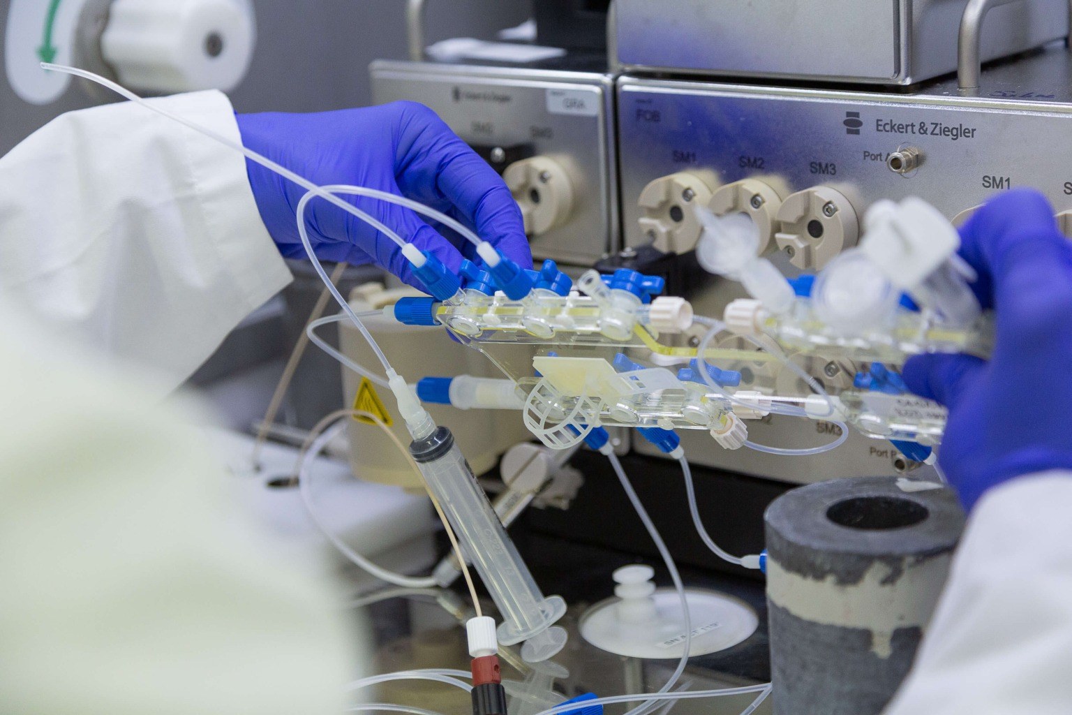
Radiopharmaceutical Chemistry includes the use of radiopharmaceuticals, which in many cases are pharmaceutical drugs, which are coupled with a molecule emitting radioactivity. Its applications can be found in diagnosis and therapy, including cancer. Cancer diagnosis with radiopharmaceuticals is coined with the term Molecular Imaging, during which we can visualize cancer in molecular/cellular level. Cancer cells have different characteristics than healthy cells, such as specific receptors on their surface. One example is the combination of the radiopharmaceutical that can recognize these receptors (-pharmaceutical) and at the same time emits a specific type of radioactivity (radio-), which can then be visualized in an image using specific instruments (PET, SPECT scanners). This is a very sensitive technique which, when used with the classic imaging techniques (CT) that give anatomical features of the patients, can yield a three-dimensional image of the tumor. This technique is noninvasive since it does not require any type of surgery. The patient is given a radiopharmaceutical and then is transferred to specific scanners to obtain the image. In the case of cancer therapy, the radiopharmaceutical contains a emits a different type of radioactivity that has the ability to kill cells. When this type of radioactivity is combined with a pharmaceutical drug that can recognize the cancer cells, then the radioactivity is transported and emitted exclusively at the tumor in a targeted manner.
Usually, people relate radioactivity with a negative feeling. Could you explain to us the ways we now use radioactivity in Nuclear Medicine?
Indeed, usually when people hear radioactivity, they automatically think of disaster, such as the one in Chernobyl. This is true, of course; however, we should not forget that radioactivity exists practically everywhere, even in the air. Radioactivity’s effects on our body work in a very complicated way: sometimes they benefit us and sometimes they harm us, depending on the type, intension, and energy of the radioactivity. The toxicology rule also finds practice in radioactivity: “The dose makes the poison”.
Nuclear Medicine is a branch of medicine that uses radioactive substances, the so-called radiopharmaceuticals, as diagnostic and therapeutic tools. The difference between the two lies in the amount of the injected dose to the patient. When used as a diagnostic tool, the amount of the radiopharmaceuticals is small. In the case of targeted therapy, the amount injected is much higher. What is very important to highlight is that before any decision regarding the injected dose per patient is made, we always weigh the possible benefit of each patient against the “danger” that he/she might be under. Additionally, thanks to very strict radiation safety measures, the possible radiation exposure for other people and the environment is eliminated.
What are the advantages of the PET and SPECT diagnosis when compared with other imaging techniques, such as MRI or CT?
The techniques used in Nuclear Medicine, which include SPECT and PET imaging, are non-invasive tools to trace, quantify, and monitor the progress of treatments and the many different biochemical processes that take place during a disease. They do so by visualizing the target. The biggest advantage is that we can visualize the target at a molecular level. That allows for the right diagnosis and, consequently, in certain cases, the treatment of pathological disorders before these disorders can alter the physiology of the patient and become observable by classic imaging techniques.
You have many publications where you use small molecules or peptides that target specific receptors. Could you explain to us the procedure that you follow to choose the receptors/targets and the molecules you use?
Choosing the target requires a full understanding of the physiological changes involved in a certain disease. Targets can be certain cellular proteins that are expressed in a specific organ, biochemical pathways that occur in cancer cells, or receptors that are expressed in cells that can be found in the blood circulation. As a result, the choice of a target can have a severe impact on the diagnosis and also in the following therapeutic choices, including the targeted therapy using radionuclides. As you see, the choice of the target is not easy, and therefore, we are in close contact with nuclear physicians so we can choose together targets that are of interest in Nuclear Medicine. When we find the biochemical pathway we wish to target, the next step is to choose the right molecule to target it.
After choosing the receptor/target and the molecule you will use, how do you choose the right radioisotope?
As we mentioned above, one of the advantages of using radiopharmaceuticals is the non-invasive diagnosis and treatment of cancer. Radiopharmaceuticals contain, apart from the biomolecule, a radionuclide- the source of radiation- which is incorporated in the biomolecule using different chemical methods. There is a wide variety of radionuclides, which are radioactive isotopes of natural elements; by choosing the right one, we can make a huge impact in Nuclear Medicine.
In general, a radionuclide emits radioactivity alpha (α), beta (b), or gamma (γ). The type of emission and the energy emitted are the main factors determining which radionuclide we use. For example, for therapeutic purposes, the radionuclide of choice must have an ideal half-life, it must emit the right type of radioactivity, and it must have an affordable production route. On the other hand, a radionuclide that will be used for diagnostic purposes must have a short half-life to ensure the minimum radiation exposure of the patient, but at the same time long enough for the diagnostic procedure.
The Gastrin Releasing Receptor (GRPr), which is overexpressed in many cancer types, has been the center of cancer research for many years. Many groups trying to develop radiopharmaceuticals to target that specific receptor are choosing peptides as biomolecules, the so-called agonists. Could you explain the reason behind that strategy?
Before I answer your question about the GRPr receptor, I think it is very important to make a short introduction to the use of agonists and antagonists in radiopharmaceutical chemistry.
Agonists are radio-labeled peptides that can be internalized in cancer cells after they attach to the receptor. They cause high accumulation of radioactivity within the cancer cell, which leads to good imaging and therapy. This is why research up to fifteen years ago focused on the development of these peptides.
Interestingly though, it was in 2006 that a study was first to show that radiolabeled peptides targeting the receptor of somatostatin and act as antagonists can give similar or even better results when compared to the agonists molecules. Antagonists are these peptides that can attach to their receptor while remaining on the cellular membrane without internalization. Since then, studies on the use of these types of peptides have given extraordinary results and have reached the clinic successfully.
The GRP receptor is overexpressed in many cancer types and, therefore, can be a useful molecular target to develop radiolabeled biomolecules (peptides). Despite the huge variety of agonist peptides synthesized and developed, only a few of them have reached the clinic because of their side effects. These issues, in combination with the successful example of radiolabeled antagonists of somatostatin, have pushed many groups toward the development of radiolabeled antagonists to target the GRP. The positive results of these groups further support the use of antagonists to target the GRP.
In one of your publications, you synthesized the radiopharmaceutical agent LF1 (GRPr-based antagonist-AAZTA) for use in Theranostics. What does Theranostics include, and how useful can LF1 be in that field?
Theranostis is a term that comes from the combination of two Greek words: therapy (θεραπεία, thera-) and diagnosis (διάγνωση, -nostics). We could say that this term reflects the transition of standard medicine towards personalized medicine. Theranostics gives an adjusted therapy window based on the uniqueness of each individual and results in the right medicine for each patient at the right time. In Nuclear Medicine and Molecular Imaging, the disease and the stage of the disease are being monitored for each patient using the appropriate radiopharmaceutical diagnostic tool. Additionally, every radiopharmaceutical product is a specific and selective molecule. In the clinic, we did that by using one molecule in which we incorporated two different radionuclides: one used for imaging and one for treatment.
To answer the second part of your question, in our publication, we designed and evaluated a peptide analogue that acts as an antagonist. In that molecule, we can incorporate many different radionuclides based on whether it will be used for diagnosis or therapy. Our goal is to combine diagnosis and targeted treatment in such a way that we can collect information on the phenotype of the cancer and treat cancer patients in a personalized manner.
Your research output includes many publications on clinical trials. What has this collaboration with physicians offered to you?
Imagine Nuclear Medicine as a triangle. The three points of the triangle are the physicians, the radiopharmacists, and the equipment needed. The patient is right at the center. Therefore, having always as our top priority the patient, the constant collaboration of radiochemists and physicians is more than a necessity. The close collaboration of radiochemists with the physicians is as crucial as it is the close collaboration with the pathologists, neurologists, and oncologists. Together with the physicians, we choose the target and, if the preclinical evaluation of the new radiolabeled pharmaceuticals looks promising, together we can plan the next step: the clinical evaluation.
Your new position is head of a new lab. How difficult can it be to build your own research group from scratch? What will be the focus of your group?
Indeed, in April of 2018, I accepted the honorable offer of the director of the Nuclear Medicine of University Medical of Bern, in Switzerland, Prof. Axel Rominger, to lead the new Radiopharmaceutical Chemistry lab. There was no such lab in this department before, so we needed to start from scratch. Even if it is the hardest project I have ever done, I can surely say that it represents one of the best lessons I have had so far.
Our group will focus on two fields: oncology and neurology. Our goal is to plan and evaluate radiopharmaceutical agents for the diagnosis and treatment of many cancer types and neurological diseases, having as an ultimate goal their translation to the clinic.
One of the most promising aspects of Radiopharmaceutical Chemistry and, therefore, of Nuclear Medicine is Personalized Medicine. Having a deeper understanding of physiological and pathological processes, we can advance towards a more accurate prediction of the treatment response a patient can show.
Radiochemistry is a relatively young field, since the first use of radiopharmaceuticals by George de Hevesy was in 1924. Today, after almost a century, how do you assess the progress of Radiochemistry? How can you predict the future of this field?
During this century, the development of radiochemistry has been rapid, and today we are in a highly advanced stage, mainly because we keep getting better at the understanding of three things:
Today, there are more than a hundred different imaging techniques, and we can perform imaging in each organ of the human body. These techniques allow the treatment and diagnosis of neurological diseases, cancer, gastrointestinal diseases, urogenital diseases, coronary artery disease, bone diseases, any injuries, infections, and lung diseases.
One of the most promising aspects of Radiopharmaceutical Chemistry and, therefore, of Nuclear Medicine is Personalized Medicine. Having a deeper understanding of physiological and pathological processes, we can advance towards a more accurate prediction of the treatment response a patient can show.
How do you think Greece is doing in the fields of research and production of radiopharmaceuticals? How do you foresee the future?
Greece has made clear progress in the research and production of radiopharmaceuticals in the last few years. When I first started my studies in radiochemistry back in 2002, there were less than ten master students and few laboratories. Nowadays, more students choose to study in our field, the laboratories in Greece have been upgraded and the country is standing out in the field. At the same time, there is a cyclotron for the production of short-lived isotopes and their distribution in hospitals across the country. We can only be proud of all these achievements and surely the future seems bright.
What would you say to someone who wants to embark on a journey in the field of Radiopharmaceutical Chemistry?
Radiopharmaceuticals is a science that combines fundamental research with an immediate impact on patient care. Our field exists right at the crossroads of medicine, biology, chemistry, and physics. In addition, the applications of Radiopharmaceutical Chemistry in Nuclear Medicine take place on an international level. Therefore, it is certainly not a dull field. We will be very happy to welcome new scientists into the fascinating world of Radiopharmaceutical Chemistry.
Follow Dr. Eleni Gourni:
Aglaia (Lina) Ntokou

Dorothea Maneta

Aglaia (Lina) Ntokou

Chrysovalantou Kalaitzidou